
 Publish:2026-10-10
 Source:ReedBiotech
 Views:85
In leading immunology journals, IL-17A and TGF-β1 are widely recognized as a key pair of immune biomarkers. Their relationship is not simply a linear upstream–downstream interaction; rather, they represent a classic interplay of cooperation and antagonism, with each contributing to the regulation of the other’s biological effects .
Measuring either biomarker alone provides only a partial picture of the inflammatory response. Combined measurement and ratio analysis offer a more comprehensive assessment of changes in immune homeostasis.
Why Are IL-17A and TGF-β1 Considered an Immune Duo with Opposing Effects?
TGF-β is a cytokine with a particularly distinctive dual regulatory role in the immune system:
High TGF-β1 levels in the absence of strong pro-inflammatory signals: TGF-β1 promotes the differentiation of naïve CD4+ T cells into regulatory T cells (Tregs), supporting anti-inflammatory responses and maintaining immune tolerance.
Low TGF-β1 levels in an IL-6-rich inflammatory microenvironment: The differentiation pathway shifts, promoting the development of pathogenic Th17 cells from naïve T cells. These cells produce substantial amounts of IL-17A, amplifying inflammation and contributing to tissue damage.
A review has highlighted that TGF-β1 is a cytokine with dual functions. It can mediate immune tolerance and maintain physiological homeostasis, but it can also contribute to pathogenic inflammatory responses. Its ultimate biological effects are determined by the dynamic cytokine milieu in the local microenvironment
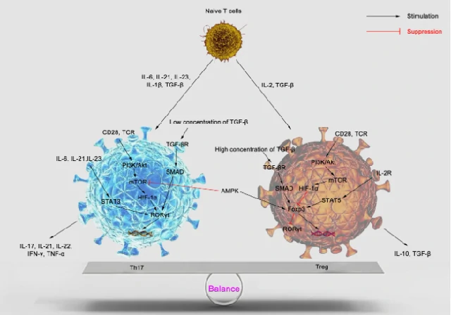
Figure: Schematic illustration of the Th17/Treg immune balance
Understanding the Th17/Treg Balance Is Key to Understanding This Immune Duo
Th17 and Treg cells both differentiate from naïve CD4+ T cells. Together, they form a finely regulated counterbalancing system within the immune system and provide the cellular basis for understanding the IL-17A/TGF-β1 biomarker pair.
Th17 Cells: The Immune System's Offensive Arm
Th17 cells are primarily characterized by their secretion of IL-17A and play an important role in host defense against infections. However, when abnormally activated, they can sustain inflammatory tissue damage and serve as key pathogenic contributors to autoimmune diseases, including rheumatoid arthritis, inflammatory bowel disease, psoriasis, and multiple sclerosis.
Studies have shown that Th17 cells are not a uniform population. They can be divided into highly pathogenic Th17 (pTh17) and less pathogenic Th17 (cTh17) subsets. The differentiation, functions, and molecular characteristics of these two subsets are precisely regulated by signaling pathways within the TGF-β superfamily.
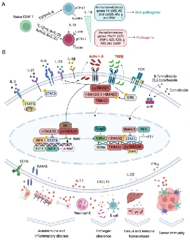
The TGF-β superfamily broadly regulates Th17 cell biology and contributes to the maintenance of physiological homeostasis and the progression of various diseases.
A. Activated CD4+ T cells can differentiate into highly pathogenic pTh17 cells or less pathogenic cTh17 cells under different cytokine conditions. These two subsets exhibit distinct molecular characteristics.
B. During Th17 differentiation, TGF-β and activin A, both members of the TGF-β superfamily, integrate signals from the T-cell receptor (TCR), interleukins, and the aryl hydrocarbon receptor (AHR). Through Smad signaling and interacting proteins, these pathways regulate the expression and activity of RORγt, thereby controlling Il17 transcription. Mature Th17 cells secrete multiple cytokines and participate in immune responses, autoimmune disease, tumor development, and the maintenance of physiological homeostasis .
Treg Cells: The Immune System's Braking Mechanism
Regulatory T cells (Tregs) are characterized by the lineage-defining transcription factor Foxp3. Through mediators such as TGF-β1 and IL-10, they suppress excessive activation of T cells, macrophages, and dendritic cells, helping prevent the immune system from attacking the body's own tissues. They also contribute to tissue repair following injury and help maintain immune homeostasis.
Immune Balance vs. Pathological Imbalance
Balanced homeostasis: Th17 cells promote inflammation, while Tregs suppress it. When these opposing functions remain balanced, immune responses are appropriately regulated.
Pathological imbalance: Abnormal Th17 activation and impaired Treg function shift the immune balance toward inflammation, contributing to autoimmune and chronic inflammatory diseases.
What the Literature Tells Us: Don't Just Track Changes—Analyze the Ratio
Many experiments focus solely on whether individual biomarkers, such as IL-17A or TGF-β1, increase or decrease. However, changes in a single biomarker may not provide sufficient evidence to support a strong conclusion. The IL-17A/TGF-β1 ratio may provide additional insight into disease activity and immune balance.
Clinical studies have reported a significantly elevated serum IL-17A/TGF-β1 ratio in patients with autoimmune diseases. Compared with measuring IL-17A or TGF-β1 alone, the ratio may better reflect the state of immune dysregulation.
Consider a drug intervention study in which only IL-17A is measured. A decrease in IL-17A might be interpreted as evidence of therapeutic efficacy. However, simultaneous measurement of TGF-β1 may reveal that TGF-β1 has also decreased. If the IL-17A/TGF-β1 ratio shows no significant reduction, the Th17/Treg immune balance may not have been effectively restored, suggesting that the intervention's overall impact on immune regulation may be limited.
This illustrates the scientific rationale for measuring both biomarkers rather than relying on either one alone.
Immune biomarkers rarely tell the whole story in isolation. Selecting the right biomarker pair and understanding the relationship between their biological functions are equally important. The IL-17A/TGF-β1 pair is relevant to a broad range of autoimmune and inflammatory research. Combining biomarker measurements with ratio analysis can help address interpretive limitations and strengthen the scientific rationale and persuasiveness of research findings.
Practical Tips for Researchers
1. TGF-β1 sample preparation
In vivo, much of TGF-β1 exists in a latent form. Before measurement, samples may require acid activation to release active TGF-β1. Follow the sample acidification and activation procedures specified in the assay kit instructions.
2. Data presentation and interpretation
Consider reporting the IL-17A/TGF-β1 ratio alongside the individual biomarker concentrations. This approach provides a more integrated view of the relationship between inflammatory and regulatory signals and may align more closely with research approaches used in high-impact immunology publications.