
 Publish:2026-10-08
 Source:ReedBiotech
 Views:95
In one sentence: The key difference lies in whether the target is bound to plasma proteins and in the different biological significance reflected by each form.
Simply put: The bound form can be regarded as a “storage” or “transport” state, whereas the free form is the “active state” that can actually exert biological effects.
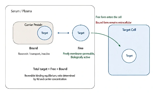
Figure 1. Free and Bound Forms of Targets in Serum: Concepts and Biological Significance
Specific Differences Between the Two Forms
- Different concepts: The free form refers to the target existing independently in the blood without being bound to proteins; the non-free (bound) form refers to the target bound to specific plasma proteins. The sum of the two is the “total” level.
- Different biological activity: The free form can freely cross cell membranes and directly participate in physiological processes, making it the fraction that actually exerts biological activity; the bound form mainly serves storage and transport functions and cannot directly enter tissues.
- Different significance for detection: The free form can more directly reflect the target’s “real-time functional state” and is not affected by changes in binding-protein concentrations; the total level reflects the overall amount secreted, but may show “false increases or decreases” due to changes in protein levels.
Common Examples
1. Thyroid Hormones (T4/T3)
Approximately 99.98% of circulating T4 is protein-bound, with only 0.02% present as free FT4; approximately 99.7% of T3 is bound, while only 0.3% is present as free FT3. Measurement of free FT4/FT3 directly reflects thyroid function, is not affected by binding-protein concentrations, and is more sensitive than total T4/T3, particularly in conditions with abnormal protein levels such as pregnancy and liver disease.
2. Prostate-Specific Antigen (PSA)
Approximately 80% of serum PSA is protein-bound, while about 20% is present in free form. When total PSA falls within the 4–10 ng/mL “gray zone,” total PSA alone is difficult to use to distinguish prostate cancer from benign prostatic hyperplasia. In this situation, calculating the free PSA/total PSA ratio is more valuable—the lower the ratio, the higher the risk of cancer (approximately 56% when the ratio is <0.1, compared with about 8% when it is >0.25).
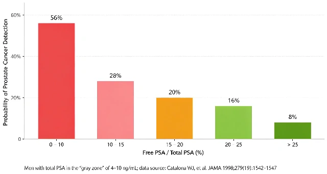
Figure 2. Free PSA / Total PSA Ratio and the Probability of Detecting Prostate Cancer (Total PSA 4–10 ng/mL)
3. Testosterone
Total testosterone includes testosterone tightly bound to sex hormone-binding globulin (SHBG) and therefore essentially unavailable for use, testosterone loosely bound to albumin and therefore releasable, as well as a small amount in completely free form.
Only free testosterone can enter cells and exert its effects. If SHBG is abnormally elevated, total testosterone may be “pulled down” and appear low, while free testosterone may remain completely normal. In this situation, blindly administering hormone replacement based on total testosterone would be the wrong approach.
4. Cell-Free DNA (cfDNA)
cfDNA consists of free nucleic acid fragments released into the bloodstream through cellular apoptosis or necrosis. It is present at very low concentrations (ng/mL range) and consists of relatively short fragments (approximately 200 bp). It has become a key biomarker in areas such as liquid biopsy for cancer and prenatal screening, placing high requirements on the sensitivity and purity of extraction reagents.
Cell-Free DNA in Peripheral Blood (Circulating Tumor DNA, ctDNA)
As a new tumor biomarker, ctDNA is increasingly playing an important role in cancer diagnosis, treatment, and prognostic assessment. It is particularly useful for tumors that lack typical clinical symptoms, have nonspecific examination findings, or are difficult to diagnose, potentially avoiding complex and invasive biopsies.
As Forshew stated, the prospects for circulating tumor biomarkers are enormous because they represent cutting-edge noninvasive testing technologies with high sensitivity and specificity and provide a high level of reliability for clinical auxiliary diagnosis.
With advances in cancer molecular biology research, plasma cell-free circulating tumor DNA testing is expected to become an important molecular biological testing approach for early cancer diagnosis, prognostic assessment, and follow-up monitoring of cancer patients.
5. What Are the Differences Between HCG, β-HCG, and Free β-HCG?
HCG: A biologically active dimer composed of α and β subunits.
Total β-HCG: The β subunit of HCG, which has high immunological specificity and does not cross-react with other hormones. It generally includes the free β subunit and HCG.
Free β-HCG: Native free β subunit and nicked free β subunit. It is often associated with trophoblastic cell disorders.
There is little difference in the clinical significance among the three. However, in terms of detection methods, total β-HCG and free β-HCG have less cross-reactivity with other proteins and therefore have higher specificity.
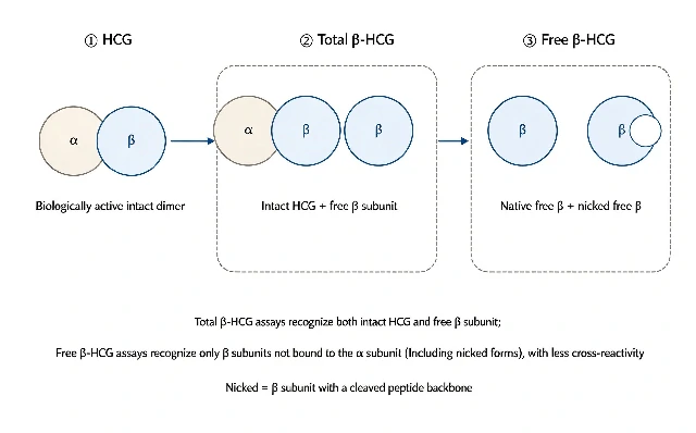
Figure 3. Molecular Forms and Recognition Differences Among HCG, Total β-HCG, and Free β-HCG
6.Urine HCG vs. Blood HCG: Which Is More Accurate?
Urine HCG: In addition to intact HCG, urine contains many free β-HCG molecules and their degradation fragments. At the same time, the concentration of HCG in urine is related to the degree of urine concentration. The more concentrated the urine, the higher the HCG concentration, as in first-morning urine. Urine HCG testing is mostly qualitative, with results determined visually, and is greatly affected by individual factors.
Blood HCG: Blood HCG testing is mostly quantitative and provides a specific numerical value. Through continuous monitoring, it can help distinguish slight increases in HCG during menstruation, nonspecific increases caused by pituitary and other disorders, ectopic pregnancy, and early miscarriage. Qualitative urine HCG testing cannot provide these distinctions. Therefore, blood HCG testing is more accurate.
One-Sentence Summary
Testing the free form of a target essentially means looking at the “fraction that can actually function,” while testing the total target means looking at the “overall amount present.” Clinically, the ratio or difference between the two is often used for more precise assessment, particularly when distinguishing benign from malignant conditions, evaluating endocrine function, and avoiding interference from binding proteins. Free-form indicators are often more informative.
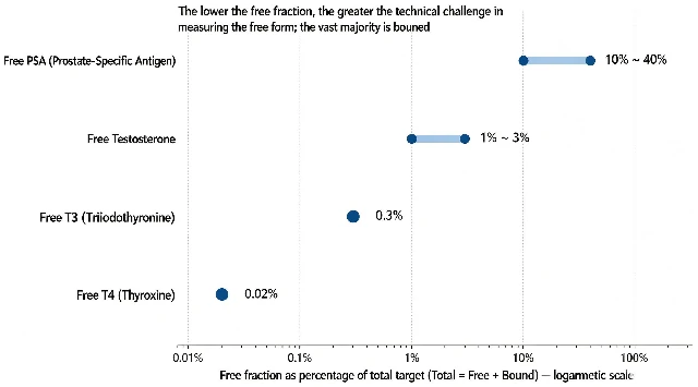
Figure 4. Proportion of the Free Fraction Among Common Serum Targets (Logarithmic Scale)