
 Publish:2026-08-14
 Source:ReedBiotech
 Views:415
——Yale University’s Reinisch team reports two complementary studies: uncovering the full pathway of non-vesicular lipid transfer and revealing the molecular mechanisms underlying Parkinson’s disease and chorea
I. Research Background: VPS13—A Key “Bridge” for Cellular Lipid Transfer
In eukaryotic cells, most phospholipids, sterols, and other lipids are synthesized in the endoplasmic reticulum (ER) and need to be efficiently transported to the plasma membrane, lysosomes, mitochondria, and other organelles. Conventional vesicular transport accounts for only part of lipid trafficking. Bridge-like lipid transfer proteins (BLTPs) are key molecular machines that mediate high-volume lipid transport across membranes independently of vesicles.
VPS13 is one of the most important subfamilies of BLTPs. Humans encode four VPS13 isoforms (VPS13A/B/C/D), and defects in their functions are directly associated with severe inherited neurological diseases:
- VPS13A mutations: Chorea-acanthocytosis, a Huntington’s disease-like disorder accompanied by abnormally shaped red blood cells
- VPS13C mutations: Early-onset autosomal recessive Parkinson’s disease and Lewy body dementia
- VPS13B/D defects: Associated with intellectual disability and peroxisomal developmental abnormalities, respectively
- Two major questions had remained unresolved:
- How does VPS13 cooperate with a membrane scramblase to distribute lipids evenly between the two leaflets of the membrane and support organelle membrane expansion?
- · How does VPS13 undergo conformational switching to precisely regulate its lipid transfer between an “autoinhibited” and an “activated” state?
In 2026, Karin M. Reinisch’s team at Yale University published two complementary cryo-EM studies in Cell and Molecular Cell. The Cell study analyzed the mechanism of the VPS13A-XKR1 complex, while the Molecular Cell study revealed the autoinhibited conformation of VPS13C. Together, the two studies provide a more complete picture of the regulatory pathway governing lipid transfer by the VPS13 family.
II. Cell Study: VPS13A-XKR1 Complex—A Coordinated Pathway for Lipid Delivery and Membrane Scrambling
Paper title: Mechanism of lipid transfer by bridge-like protein VPS13A and the scramblase XK
Core technologies: Near-atomic-resolution cryo-EM at 3.28–3.41 Å + all-atom and coarse-grained molecular dynamics (MD) simulations
Overall mechanism of the VPS13A-XKR1 complex
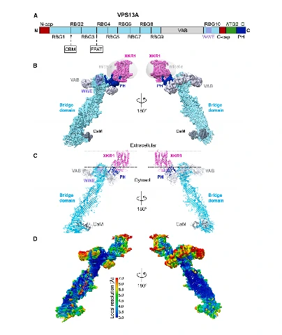
Figure 1. Global working model of the VPS13A/CaM-XKR1 complex: the cryo-EM reconstruction is shown on the left, while molecular dynamics simulations on the right reveal the spatial separation between lipid delivery and lipid scrambling.
1. Complex assembly: PH domain-mediated specific binding
VPS13A specifically binds the plasma membrane scramblase XKR1 (XK) through its C-terminal PH domain. The WWE and VAB domains are not involved in this interaction. Two loops of the PH domain insert into a conserved cytoplasmic pocket of XKR1, anchoring VPS13A to the plasma membrane surface.
Calmodulin (CaM) binds to the N-terminal bridge region of VPS13A and regulates the association of VPS13A with the ER, which serves as the lipid donor membrane.
VPS13A结构域与冷冻电镜密度
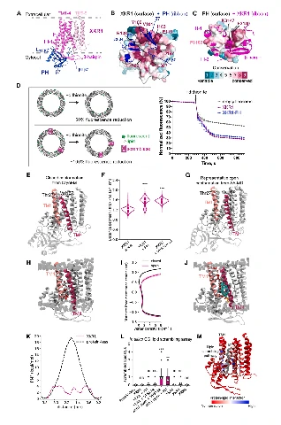
Figure 2. Domain map and cryo-EM three-dimensional reconstruction of VPS13A, showing the bridge domain and the PH/WWE/VAB adaptor modules.
2. The bridge domain directly delivers lipids to the plasma membrane
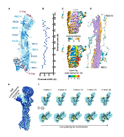
The core “bridge domain” of VPS13A forms a tube-like structure resembling a taco shell, with a hydrophobic groove inside that can accommodate dozens of phospholipid molecules and serve as a long-distance lipid transfer channel.
- The width of the channel dynamically varies along its axis from 9–25 Å, allowing only 1–2 lipids to pass side by side.
- Lipid saturation, driven by continuous lipid synthesis in the ER, provides the driving force for transfer. When the bridge domain is loaded with ≥85 lipids, the C-terminal cap undergoes a conformational opening, allowing lipids to be directly released into the cytoplasmic leaflet of the plasma membrane.
- The amphipathic ATG2_C helix of VPS13A inserts into the membrane, inducing local membrane bending and reducing the energy barrier for lipid insertion, thereby accelerating lipid delivery.Lipid Binding to the Bridge Domain and VAB Conformational Changes
3. Activation mechanism of the XKR1 scramblase: regulation by membrane tension
In the cryo-EM structure, XKR1 adopts a closed and inactive conformation. Molecular dynamics simulations show that two conditions can open the gap between TM1 and TM4:
- Binding of the VPS13A PH domain
- Increased plasma membrane tension
The difference in membrane tension between the relatively low-tension ER and the higher-tension plasma membrane generates a lipid transfer potential.
In the open conformation, a polar and hydrophilic pathway forms between TM1 and TM4, allowing newly delivered lipids to move from the cytoplasmic leaflet to the extracellular leaflet and thereby maintaining balanced growth of the two membrane leaflets.
Key findings: The lipid delivery site and lipid scrambling site are spatially separated. Their functions are independent, while both are coordinated by membrane tension.
Closed and open conformations of XKR1 and lipid scrambling simulations
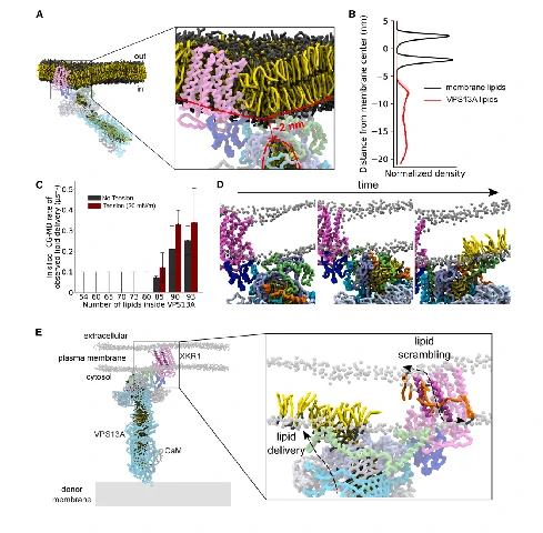
Figure 4. Closed cryo-EM conformation of XKR1, MD-derived open conformation, and free-energy simulations of lipid scrambling.
Four key highlights of the Cell study
1. The first near-atomic-resolution structure of a VPS13-scramblase complex, identifying the PH domain as the specific membrane-receptor-binding domain.
2. VPS13 directly releases lipids into the plasma membrane, without requiring the scramblase as an intermediate lipid-transfer site.
3. Lipid overload and membrane tension act as dual driving forces regulating high-volume lipid transfer.
4. A common functional model for VPS13 and ATG2 lipid-transfer proteins is established.
III. Molecular Cell Study: The Autoinhibited Conformation of VPS13C—A Molecular Switch for Lipid Transfer
Paper title: Cryo-EM structure of soluble VPS13C suggests its regulation by a conformational switch and by calmodulin
Study subject: The soluble, cytoplasmic autoinhibited conformation of the Parkinson’s disease-associated protein VPS13C, revealing the “blocking switch” function of the VAB domain and the regulatory role of calmodulin (CaM).
Global model of VPS13C activation and autoinhibition
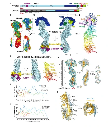
Figure 5. VPS13C regulatory model: cytoplasmic VPS13C remains autoinhibited under resting conditions and undergoes a conformational switch to activate lipid transfer following lysosomal damage.
1. Cytoplasmic VPS13C adopts a “lipid-transfer-off” autoinhibited state
The cryo-EM structure of purified, soluble VPS13C shows that its C-terminal VAB domain arches across the end of the bridge domain and completely blocks the lipid-release exit, forming an inactive lipid-transfer conformation.
The VAB domain interacts tightly with the C-terminal cap of the bridge domain through multiple hydrophobic interactions, physically preventing the bridge domain from contacting the target membrane and avoiding uncontrolled lipid release by VPS13C in the cytoplasm.
Close-up View of the VAB Domain Blocking the Bridge Domain
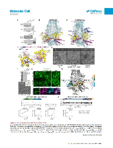
Figure 6. Close-up view showing the VAB domain covering the end of the bridge domain and blocking the lipid-output channel.
2. Lysosomal damage triggers a conformational switch and releases autoinhibition
VPS13C localizes to ER-lysosome membrane contact sites. When the lysosomal membrane is damaged, Rab7, a lysosomal small GTPase, together with the ATG2_C amphipathic helix at the membrane-defect region, promotes a conformational change:
- The VAB domain moves from the end of the bridge domain to its side, releasing the lipid exit.
- The C-terminal cap becomes flexible and opens.
- The end of the bridge domain associates with the lysosomal membrane.
This generates an activated lipid-transfer conformation consistent with that observed for VPS13A-XKR1.
Complete Regulatory Pathway of VPS13C
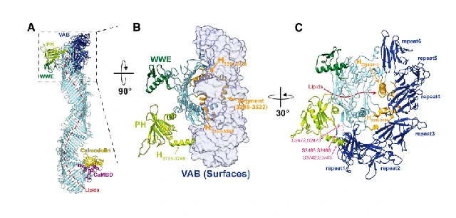
Figure 7. Complete regulatory pathway of VPS13C from autoinhibition to activation.
3. Calmodulin is an essential partner for VPS13 folding and localization
This study demonstrates that CaM forms stable complexes with VPS13A/C/D, but not VPS13B, and binds to the N-terminal region of the bridge domain.
- In the absence of CaM, VPS13 undergoes abnormal folding and cannot form its functional rod-like structure.
- VPS13 mutants defective in CaM binding cannot be recruited to ER-lysosome membrane contact sites.
- Calcium-bound CaM induces local conformational changes that fine-tune the conformation of the bridge-domain channel and regulate lipid-transfer efficiency in response to calcium signals.
Key innovations of the Molecular Cell study
1. The full-length autoinhibited conformation of VPS13 is resolved, revealing the VAB domain as a natural “off switch” for lipid transfer.
2. CaM is identified as a conserved molecular chaperone of the VPS13 family, regulating protein folding and organelle localization.
3. A three-component activation pathway involving lysosomal damage, Rab7, and the membrane amphipathic helix is revealed.
4. The two studies together establish a complete regulatory logic for the VPS13 family: autoinhibition → membrane-receptor binding → lipid release → membrane leaflet equilibration.
IV. Integration of the Two Studies: A Unified Working Model for the VPS13 Family
By combining the activated VPS13A structure with the autoinhibited VPS13C structure, the researchers propose a general lipid-transfer process applicable to human VPS13 proteins:
Resting autoinhibited state
Cytoplasmic VPS13 binds CaM to maintain its proper folding, while the VAB domain blocks the end of the bridge domain, preventing lipid release.
Target-membrane anchoring and activation
VPS13 binds membrane proteins through its PH domain, including membrane scramblases such as XKR1/ATG9, while the ATG2_C helix inserts into the target membrane. Rab7 and other GTPases help move the VAB domain away, releasing the blockade of the lipid-transfer channel.
High-volume lipid delivery
Continuous lipid synthesis in the ER fills the VPS13 bridge domain. Lipid overload drives opening of the C-terminal cap, allowing lipids to be directly released into the cytoplasmic leaflet of the target membrane. Increased membrane tension further promotes lipid release.
Membrane leaflet equilibration
The membrane scramblase XKR1 opens in response to membrane tension and transfers lipids from one leaflet to the other, supporting membrane expansion and repair.
The two studies complement each other: VPS13C explains how lipid transfer is kept “off” under resting conditions, while VPS13A explains how lipid transfer and membrane equilibration are activated after VPS13 engages with membrane proteins. Together, they connect the entire process from the cytoplasmic resting state to membrane growth.
V. Disease and Clinical Significance
1. Chorea-acanthocytosis — VPS13A mutations
Disruption of the interaction between VPS13A and XKR1 affects plasma membrane lipid transfer and phosphatidylserine scrambling, leading to disruption of neuronal membrane homeostasis and causing motor abnormalities.
2. Early-onset Parkinson’s disease — VPS13C mutations
Mutant VPS13C may fail to bind CaM or respond appropriately to lysosomal damage signals. Impaired lipid repair of the lysosomal membrane disrupts lysosomal degradation function and ultimately contributes to the death of dopaminergic neurons.
3. Potential common therapeutic targets
The CaM-binding pocket, VAB conformational regulatory sites, and the PH-XKR1 interaction interface may serve as potential targets for small-molecule drug design. Modulating VPS13 lipid-transfer activity could potentially help alleviate neurodegenerative diseases.