1. How to avoid hemolysis during blood collection?
The main causes of hemolysis:
① Mechanical damage: too slow or too fast blood collection speed, violent mixing, too thin needle (<22G), repeated blood collection;
② Improper operation: blood collection before alcohol is completely evaporated, excessive negative pressure during blood drawing, strong impact force when blood is injected into the collection tube;
③ Sample separation and preparation: prolonged processing, excessive centrifugal force or repeated freezing and thawing of samples;
④ Anesthesia blood collection: use of hemolytic anesthetics;
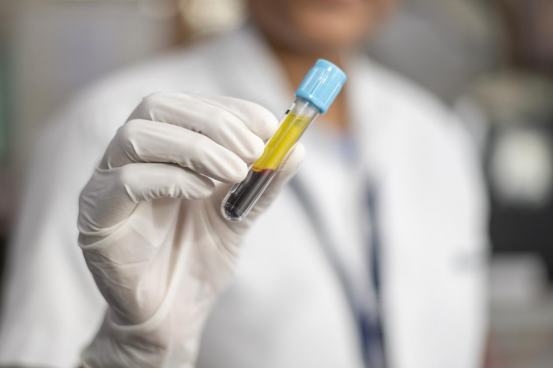
Figure 1: The normal serum plasma in the yellow part of the supernatant
① Before blood collection: Select an appropriate blood collection needle, and ensure that the alcohol has completely evaporated before collecting the blood; if performing anesthesia blood collection, it is recommended to use an anesthetic without hemolytic effect;
② During blood collection: Keep the needle stable, avoid adjusting the needle position within the blood vessel, to prevent red blood cell rupture; Control the aspiration speed, avoid forcefully pulling the syringe, and allow the blood to flow naturally;
③ After blood collection: Gently remove the needle, slowly inject the blood along the wall of the test tube, avoid bubbles and shock force; Immediately gently invert and mix the collection tube 5-8 times, and prohibit shaking;
④ Sample preparation: Process the samples according to the recommended temperature and speed, avoid prolonged time or excessive centrifugal force causing hemolysis; Try to avoid repeatedly freezing and thawing the samples;

Figure 3: Precautions for Blood Collection
2. Why is it not recommended to use hemolyzed samples in ELISA experiments? If hemolysis occurs, are there any salvage methods?
The hemolyzed samples will release endogenous HRP enzymes and hemoglobin, which have similar HRP activity and non-specifically bind to biotinylated antibodies, resulting in uncontrollable non-specific color development in ELISA and interference with the accuracy of the experimental results.
Hemolysis is classified as the following situations:
① The sample appears pink when viewed with the naked eye, this is mild hemolysis;
② The sample appears red or dark red when viewed with the naked eye, this is severe hemolysis;
③ The sample appears blackish red when viewed with the naked eye, this is oxidative severe hemolysis;
For the first situation, the supernatant can be collected by centrifugation again. For the second and third situations, it is recommended to collect the sample again!